


The valgus deformation of the foot is usually congenital. However, in some cases - with paralysis, traumatic lesions - may have appeared in mature life periods. The main symptoms of pathology are soreness in the muscles of the feet and lower legs, visible invasion of the feet, and changes in gait. Clinical examination, X-ray, electromyography, etc. are used to diagnose the disease. Treatment includes conservative and surgical approaches. However, only appropriate validity was observed in the reconstruction operation.
What disease is this?
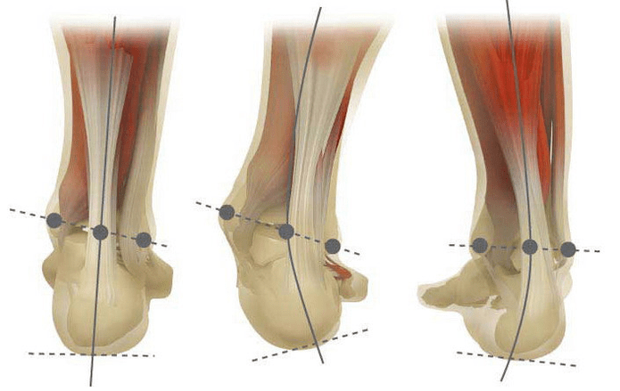
The peripheral deformation is the curvature of the foot, characterized by its longitudinal flatness. Usually, the inner edge of the foot is lowered ("drip") while the heel is lowered.
A person's feet, by virtue of their position, are subject to pressure from the entire human body. Therefore, it has a special anatomy that can depreciate, balance and stabilize movement. However, an important part of achieving these tasks is the correct stop form.
Today, the most important issue in trauma and orthopedics is the valgus deformation of the foot. The conference estimated to be 30-58%, with 2/3 of the cases constitutive diseases.
This pathology is largely socially significant because it covers all ages in all populations and also contributes to curvature of the spine, early development of osteochondrosis, and arthritis in the lower limb joints.
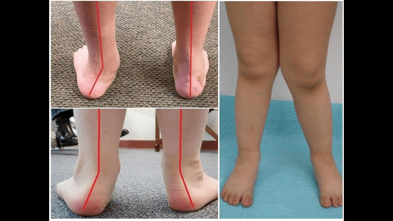
When you step on your feet (if you look at them from behind), the ankle level creates an X-shaped deformation: the ankle is in contact, while the distance between the heels is 5-6 cm.
Most of the time, pathology is inherently congenital and is diagnosed in children in hospitals (or immediately after the onset of the walk). After adjusting for up to 5 years of similar conditions, the child will develop flat feet afterwards (without proper treatment).
Why does it appear?
It is believed that the main reason for the valgus deformation of the foot is insufficient function of the posterior tibial muscle or the weakness of the ligament instrument.
Today, other factors in pathological development have been distinguished:
- Congenital abnormalities, incorrect position of the bones in the foot or shortening of tendons (vertical rod bones, short threaded tendons);
- When the foot deforms, posture disorders compensate for the curvature of the spine;
- Traumatic lesions (fractures of the feet, calf, hip or knee, rupture of ligaments and tendon devices);
- Paralysis caused by neurological damage, encephalitis, polio, stroke, myelitis of the myelitis of the myelitis of the brain, paralysis (fixation);
- Spasm of lower leg muscles (constant contraction);
- Concomitant diseases: pathology of the bone system with vitamin D (RICKET), diabetes, osteoporosis (reduction of bone density), damage to the thyroid and parathyroid glands, etc.
- Weight gain, including rapid weight gain during menopause or during pregnancy.
The development of pathology is also facilitated by overcorrection of missed shoes or club poles during childhood.
The degree and stage of the disease
The severity of pathology (the power of expressiveness) is divided into degrees:
- The vault height is 1. 5-2 cm, and the heel is tilted to 15 degrees of light;
- Average, when the arch flattened to the first centimeter, the angle dropped to 10 degrees;
- At a height of up to 0. 5 cm, the heel is inclined at 5 degrees.
Depending on the participation of certain structures, the following stages of curvature are distinguished:
- No bone deformation, and pain is determined on the inner surface of the ankle (in the fixation area of the posterior tibial muscle);
- The bend is light and the heel is somewhat rejected.
- Distribute the foot and fix the deformation (not corrected);
- Stasis is observed not only on the feet, but also in the ankle joint.
symptom
In the first stage, after a prolonged walking or a prolonged vertical load (standing or sitting on the feet), the patient is disturbed by periodic pain. Often, pain syndrome is exacerbated when walking in improperly selected shoes. The next stage of the disease is related to the occurrence of foot curvature: the patient in the standing position does not rely on the outer edge of the foot, but is related to the entire area. A slight change in gait.
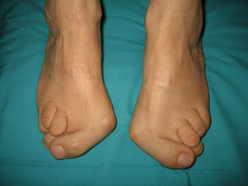
In the third stage, the protrusion of the thawed bone (significantly lower than the ankle joint on the inner surface of the ankle), and the strong metastasis of the external heel (the patient is based on the inner edge of the heel bone). Advanced valgus deformation of the foot is characterized by the obvious curvature of the foot itself and the ankle joint. The patient complained about severe pain in the lower leg muscles and severe gait violations: the knees rub against each other, while the left foot and left foot are at a distance.
Deformation of the spine of the foot (the wing position of the shoulder colo bone and pelvis in scoliosis is different), osteocartilage changes (damage to the disc, forming a hernia) or arthritis (injury within the periosteum) or arthritis (in the lower calf and hip rod), usually due to different positions of the wings of the shoulder and bone (damage to the disc damage).
How to diagnose?
Diagnosis of foot bend includes:
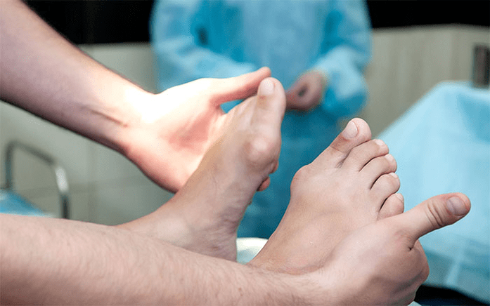
- During this clinical examination, orthopedics detected a decrease in the arch of the foot, deviation of the heel and bone, visible "disappearance" of the external ankle, and protrusion of the internal ankle.
- x -ray - An affordable and informative method where you can determine the change in its relationship by the angle of its bone inclination and linear parameters. These indicators are necessary to make the final diagnosis and clarify the degree of deformation.
- The method of registration steps is designed to determine the exact functional status of the limb. The method includes registering the support time of each part of the foot when performing the step. During the study, the phase of foot rolling was also studied, which reflects the balance of lower limb muscles.
- Dynamic electromyography records the study of electrical activity of muscles and their dependence on the step phase.
- Optical computing with digital processing enables you to obtain all standard metrics and determine the type/degree of curvature with high accuracy.
Additional consultation with a neurologist (deformation caused by spasms or paralysis), an endocrinologist (in the case of diabetes or disease of the thyroid/parathyroid gland) and a gynecologist (when worsening may be required). If the curvature of the foot occurs in the context of osteoporosis, optical density-bone density studies are required.
treat
Among the main methods for treating foot curvature, there are differences in conservatism and operation. Don't destroy joints with ointment and injections!
A conservative approach
This help is designed to get rid of the symptoms of the disease, but it does not eliminate the underlying cause of the pathology.
This technology includes:
- Use orthopedic insoles to support I Plus bones, arches of the feet and eliminate lateral lesions in the back and back of the feet;
- Tape - Use appropriate elasticity to secure your feet and ankles with special tape. The tape is worn on the clock within 3-5 days and is replaced later.
- Sew orthopedic shoes to a single standard;
- Use orthotics and other fixing devices on your feet and ankles.
Conservative approaches also include physical therapy procedures (Ozokerite, paraffin applications, electrophoresis, magnetic effects), massage and physical therapy exercises developed for specific clinical cases. Beware! Today, most experts prefer surgical treatments because conservative treatments are ineffective (as statistics, useless in 60% of cases).
Surgical intervention
The amount of operation and its type depend on the direct stage of the disease. Therefore, return to the anatomically corrected position by synovectomy (removing the tendon shell to correct general tension) or osteotomy (anatomy) of the heel. In the second stage of disease development, a transplant of finger tendon bending was used. This intervention is usually performed in the anatomical context of heel or ramming joint fixation (surgical fixation of the joint between RAM and Scaphoid Bones).
The curvature of degree III requires immediate joint fixation: several joints of five feet: without five cups and RAM. The anatomy of the heel bone can usually supplement these three reinforcement immobilizations. During the IV stage of pathology, a reconstruction operation is required not only on the foot but also on the ankle. In this case, use a graft (from its own body or artificial substance) to adjust the instability of the ligament instrument. The amount of operation on the foot is the same as the degree of curvature III.
Recovery period
Rehabilitation includes walking without supporting legs within 2 months. At the same time, the patient needs to wear movable plaster filaments within 1. 5 to 3 months. It is recommended to start active exercise 1. 5 months after the operation. By the third month, a comprehensive body for strengthening physical education was introduced. However, patients are subsequently prohibited from jogging walking and moving activities. It is worth noting that the final result of the operation can be judged only after six months.
Preventive measures
Preventing the deformation of the shell includes the following measures:
- Early correction for congenital abnormalities, inappropriate bones in foot bones or shortening of tendon grains (vertical melted bones, short heel tendons);
- Correct posture diseases (scoliosis, etc. );
- Treat traumatic lesions (fractures of foot bones, lower legs, thighs or knee joints, ligaments and tendons ruptures);
- Due to nervous system damage, encephalitis, polio, stroke, cerebrospinal roots that penetrate the roots of hernia, etc. , the paralysis will be properly recovered (fixed).
- Reduce spasms in lower leg muscles (continuously decreasing);
- Treatment of concomitant diseases: pathology of the bone system (rick bone), diabetes, osteoporosis (reduced bone density), impaired thyroid and parathyroid function, etc. ;
- weight drops to normal (especially after menopause or speeds up due to pregnancy);
- Choose orthopedic shoes or use substitute shoes;
- No "super correction" treatment is required, resulting in moderate correction of foot training for foot auxiliary surgery.
Preventing disease progression is done using conservative methods and early reconstruction operations. In this case, physical activity is limited to prevent damage and curvature of the ankle joint. Remember that prompt treatment of valgus deformation of the foot can not only improve the patient's quality of life, but also prevent the development of osteochondrosis and arthritis in the knee or hip joints!